 , Yangzi Jiang 1,2,3,7,8,*
1 Institute for Tissue Engineering and Regenerative Medicine, The Chinese University of Hong Kong, Hong Kong SAR, China
2 Department of Orthopaedics & Traumatology, Faculty of Medicine, The Chinese University of Hong Kong, Hong Kong SAR, China
3 Center for Neuromusculoskeletal Restorative Medicine (CNRM), The Chinese University of Hong Kong, Hong Kong SAR, China
4 National Clinical Research Center for Geriatric Disorders, Xiangya Hospital, Central South University, 410008 Hunan, Changsha, China
5 Department of Endocrinology, Xiangya Hospital, Central South University, 410008 Hunan, Changsha, China
6 Department of Orthopaedic, Xiangya Hospital, Central South University, 410008 Hunan, Changsha, China
7 School of Biomedical Sciences, The Chinese University of Hong Kong, Hong Kong SAR, China
8 Key Laboratory for Regenerative Medicine, Ministry of Education, School of Biomedical Sciences, Faculty of Medicine, The Chinese University of Hong Kong, Hong Kong SAR, China
Abstract
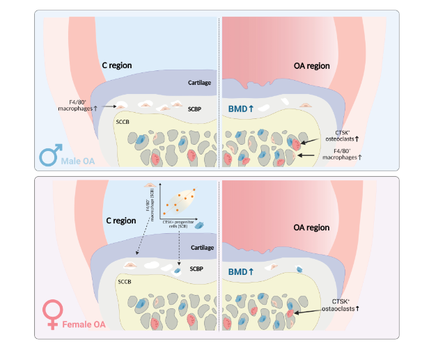
Objective: The load-bearing structures of the subchondral bone undergo alterations in osteoarthritis (OA) joints and exhibit distinct bone remodelling properties. This study examined the pathological features and cellular components of the subchondral bone plate (SCBP) and subchondral cancellous bone (SCCB) in OA-affected regions of human knee joints. Methods: Tibial plateaus were obtained from patients with varus knee OA (n = 42; women: n = 22, aged 57–87 years; men: n = 20, aged 59–82 years). Osteochondral specimens were collected from OA lesion sites in the medial compartment (OA region) and paired control sites in the lateral compartment (C region). Bone mineral density (BMD) was evaluated using micro-computed tomography, osterix+ osteoprogenitors, cathepsin K (CTSK)+ osteoclasts, and F4/80+ macrophages were quantified by immunohistochemistry, and correlations between cellular components were analysed by sex and region. Results: The OA SCBP had a significantly higher BMD than did the C region. In male patients, more F4/80+ macrophages were present in the SCBP C region than in the OA region. Female OA SCCB samples showed an increased number of CTSK+ osteoclasts. In both sexes, compared with the C region, the OA SCCB contained more CTSK+ osteoclasts and macrophages. Positive correlations between macrophage and osteoprogenitor densities were observed in most subchondral bone regions, except in male OA samples. Conclusions: Region-specific differences in cellular components were identified in the OA subchondral bone. Asynchronous remodelling responses were noted between the SCBP and SCCB. These findings provide detailed insights into OA pathology and can inform future therapeutic strategies.
Graphical Abstract
Keywords
- Macrophage
- sclerosis
- subchondral bone plate
- tibial plateau
- trabeculae
- osteoclast