eCM (Eur Cell Mater / e Cells & Materials) eCM Open Access Scientific Journal
 ISSN:1473-2262         NLM:100973416 (link)         DOI:10.22203/eCM

2018   Volume No 36 – pages 30-43

Title: Nondestructive fluorescence lifetime imaging and time-resolved fluorescence spectroscopy detect cartilage matrix depletion and correlate with mechanical properties

Authors: AK Haudenschild, BE Sherlock, X Zhou, JC Hu, JK Leach, L Marcu, KA Athanasiou

Address: University of California Irvine, 3418 Engineering Hall, Irvine, CA 92697, USA

E-mail: athens at uci.edu

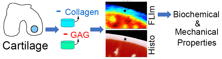
Abstract: Tissue engineers utilize a battery of expensive, time-consuming and destructive techniques to assess the composition and function of engineered tissues. A nondestructive solution to monitor tissue maturation would reduce costs and accelerate product development. As a first step toward this goal, two nondestructive, label-free optical techniques, namely multispectral fluorescent lifetime imaging (FLIm) and time-resolved fluorescence spectroscopy (TRFS), were investigated for their potential in evaluating the biochemical and mechanical properties of articular cartilage.
Enzymatic treatments were utilized to selectively deplete cartilage of either collagen or proteoglycan, to produce a range of matrix compositions. Samples were assessed for their optical properties using a fiber-coupled optical system combining FLIm and TRFS, their biochemical and mechanical properties and by histological staining. Single and multivariable correlations were performed to evaluate relationships among these properties.
FLIm- and TRFS-derived measurements are sensitive to changes in cartilage matrix and correlate with mechanical and biochemical assays. Mean fluorescence lifetime values extracted from FLIm images (375-410 nm spectral band) showed strong, specific correlations with collagen content (R2 = 0.79, p < 0.001) and tensile properties (R2 = 0.45, p = 0.02). TRFS lifetime measurements centered at 520 nm (with a 5 nm bandwidth) possessed strong, specific correlations with proteoglycan content (R2 = 0.59, p = 0.001) and compressive properties (R2 = 0.71, p < 0.001).
Nondestructive optical assessment of articular cartilage, using a combination of FLIm- and TRFS-derived parameters, provided a quantitative method for determining tissue biochemical composition and mechanical function. These tools hold great potential for research, industrial and clinical settings.

Key Words: Cartilage, biomechanics, extracellular matrix, collagens, proteoglycans, imaging, cartilage repair and regeneration, tissue engineering, regenerative medicine.

Publication date: July 27th 2018

Article download: Pages 30-43 (PDF file)
DOI:
10.22203/eCM.v036a03

Twitter Facebook Google LinkedIn Print