Microstructural morphology and mechanical analysis of decellularized ligament scaffolds
 , H. Z. Chen 1,*
1 State Key Laboratory of Oral Diseases & National Center for Stomatology & National Clinical Research Center for Oral Diseases, West China Hospital of Stomatology, Sichuan University, 610041 Chengdu, Sichuan, China
Abstract
Background: The limited self-repair capacity of ligaments imposes substantial obstacles to effective tissues healing, thereby highlighting decellularization as a viable approach for ligament reconstruction. While whole tissue decellularization retains gross morphological features, its impact on microstructural organization and multiscale mechanical behavior remains insufficiently elucidated. Methods: Rabbit medial collateral ligaments were decellularized using a combined protocol of physical, chemical, and biological methods, comprising five freeze-thaw cycles (physical), 0.38 g/mL guanidine and 0.5 % sodium dodecyl sulfate (SDS) (chemical), and 0.5 % trypsin (biological). Decellularization efficacy was validated through hematoxylin-eosin (HE) and 4′,6-diamidino-2-phenylindole (DAPI) staining and biochemical quantification of deoxyribonucleic acid (DNA), glycosaminoglycan (GAG), and collagen content. Structural and mechanical alterations across scales were assessed via scanning electron microscopy (SEM), fluorescence collagen hybridizing peptide (F-CHP) staining, atomic force microscopy (AFM), uniaxial tensile testing, and nanoindentation. Statistical analyses were performed using unpaired t-tests. Results: The decellularization process significantly reduced DNA content (from 1110.77 ± 46.16 ng/mg to 38.60 ± 1.67 ng/mg) and disrupted collagen organization, as reflected by a decreased tensile modulus (3.16 ± 0.19 MPa vs. 3.84 ± 0.23 MPa). Microscopic evaluations revealed structural alterations in collagen fibrils, including increased porosity and expanded D-banding periodicity. Nanoindentation and AFM results indicated a decrease in mechanical properties at micrometer and nanometer scales, with compromised energy dissipation capacity. Under extreme tensile conditions, decellularized ligaments demonstrated pronounced fiber tearing and misalignment. Conclusions: Decellularization induces hierarchical disorganization of ligaments matrix at both micrometer and nanometer scales, typified by collagen fiber loosening, augmented interfibrillar spacing, and expanded D-banding periodicity. These alterations collectively lead to impaired micromechanical integrity and reduced energy dissipation. The findings highlight critical structural determinants of mechanical function and provide insights for optimizing decellularization strategies to preserve microscopic architecture and meet the demands of load-bearing applications.
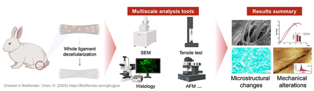
Graphical Abstract
Keywords
- Decellularization
- microstructural morphology
- mechanical behavior
- microscopic analysis
- ligaments